Picturing the Molecules of Life
- By Rebecca Deusser
- Posted 04.22.03
- NOVA
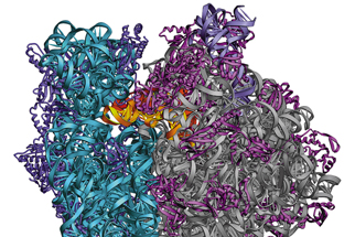
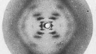
From Watson and Crick to today's molecular biologists, researchers rely on X-ray diffraction images to deduce information about of the molecules of life. Where once these images were blurry and black and white, today X-ray diffraction specialists feed data into computer programs that generate colorful 3-D images. As the technology has improved, scientists have also tackled ever more complex biochemical structures, from RNA to ribosomes. The resulting images reveal clues about their structures, their interactions with other molecular structures, and their role in human genetics. Here, see some of the most significant images of the past century.
 Launch Interactive
Over the past 50 years, scientific images of DNA, ribosomes, and RNA have catalyzed our understanding of biology.
This feature originally appeared on the site for the NOVA program Secret of Photo 51.
Credits
Photos
- (1952)
- © Franklin, R. and Gosling, R.G./Nature
- (1973, 1984)
- Courtesy of Shana Kelley, Boston College
- (1979, 1980)
- © Protein Data Bank
- (2001)
- © Center for Molecular Biology of RNA, UC-Santa Cruz
Related Links
-
Anatomy of Photo 51
Explore Rosalind Franklin's famous x-ray image, a key to understanding the double-helix structure of DNA.
-

Before Watson and Crick
How did scientists discover that DNA was the blueprint of life? Brenda Maddox explains.
You need the Flash Player plug-in to view this content.