
We recommend you visit the interactive version. The text to the right is provided for printing purposes.
|


|
|
Intro
In 2005, Mary Schweitzer and her
team shocked the paleontological world when they reported, in the journal Science, that they had come upon soft tissue surviving deep
within the fossilized thigh bone of a 68 million-year-old Tyrannosaurus
rex. The tissue included transparent,
stretchy, and still-soft structures that looked like blood vessels, along with
possible red blood cells. Surprised as anyone by the discovery, Schweitzer (left), a
paleontologist and research curator at North Carolina State University, decided
she had to see if it was a fluke. Did other ancient fossils harbor such prizes?
Here, see what Schweitzer and her team brought to light from bones as old as 78
million years.—Peter Tyson
Note: We focus on the apparent
blood vessels here, but Schweitzer's team found other soft tissues,
including bone-building cells and fibrous bone matrix. For the full story, see
her team's 2005 and 2007 papers on the findings, referenced in Links &
Books.
|
|
|
Ostrich
Present day
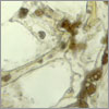
To put their work in the broadest
possible context, Schweitzer's team worked with samples from a wide range
of animal types, fossil deposits, continents, and time periods. Shown here are blood vessels from the bones of an ostrich. The
ostrich was chosen because it's one of the most primitive of living
birds, which most paleontologists now agree are the dinosaurs' closest
living relatives. It is also big and must stand on two legs, so it has similar
biomechanical constraints as some dinosaurs. The dark red blobs filling
the vessels, which weave through white bone matrix, are blood breakdown
products.
|
|
|
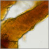
Emu
Present day
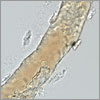
A second species they tested
was emu, the largest living bird after the ostrich and also flightless and
"primitive." This specimen had been
buried for about two years when its leg bones were exhumed for the study. As
with all its other specimens, the team dissolved the skeletal remains
in a weak acid that slowly eats away bone, including fossilized bone, but not
soft tissues. They were left with translucent blood vessels (left). This vessel's light pigment is thought to be compounds derived
from hemoglobin, which transports oxygen in the blood and lends red blood cells
their color.
|
|
|
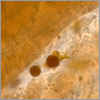
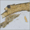
Moa
800-1,000 years old
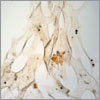
The final bird the team studied was
the moa, a giant flightless bird that lived in New Zealand until
it went extinct about 500 years ago. Resembling the roots of a tree, the clear,
interconnecting blood vessels seen here remained after this specimen's
acid bath. The small black dots are fungal spores that infiltrated the bone,
probably after the bird's death. The fact that blood vessels survived in
this and in the far older samples to follow flies in the face of conventional
thought, which holds that all soft parts degrade and disappear very quickly
after death.
|
|
|
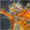
Mammoth
300,000 years old
Going further back in time, the
team tested the skeletal remains of a Columbian mammoth from the Pleistocene.
The hollow, orange-tinged vessels seen here are still flexible,
while those in the inset are crystalline and shatter easily, even though both
came from the same specimen. Why the discrepancy in fossilization? Further studies now needed to
update our understanding of the fossilization process may help tell. The main image also
shows possible red blood cells. The white material is bone matrix that
didn't dissolve in the acid—probably collagen, a chief component of
bone and connective tissue.
|
|
|
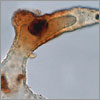
Mastodon
300,000 years old
The blood vessels found within the
bones of this American mastodon were, as seen here, mostly
crystalline—that is, gone to fossil and not soft. Only small fragments of
still-elastic vessels were uncovered. But like many of the other specimens, the
vessels were richly colored with possible blood products. Schweitzer and her
team have already speculated on how tissues might have survived for so long,
though the presence of soft vessels, the most easily degraded of tissues,
remains, they admit, "enigmatic." This bone also featured a
significant amount of preserved collagen matrix.
|
|
|
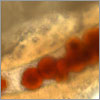
Tyrannosaurus rex
65 million years old
The team examined a number of T.
rex specimens, several of which revealed
what Schweitzer cautiously terms "round, red microstructures," as
vividly pictured here. Further tests will determine whether they are truly red
blood cells from the animal, or if they are some remnant of the fossilization
process that mimics them. If they are cells, could DNA have survived within?
Again, further tests should tell, though most experts agree it's a long
shot. The yellow material surrounding the vessel seen here is fibrous bone
matrix containing bone cells.
|
|
|
Tyrannosaurus rex
68 million years old
This specimen is the same T. rex as that reported on in Schweitzer's 2005
study. Hollow, translucent, and still-supple blood vessels were common in this
specimen. The vessel shown here has more of those "round, red
microstructures" and is distinguished by a particularly thick vessel
wall. Since DNA is presumed to be fragile—the oldest yet discovered is
from Neanderthal fossils roughly 50,000 years old—Schweitzer's team
has so far focused on finding proteins, which are more durable. Recently they
announced they had found protein in a T. rex specimen, in the form of collagen.
|
|
|
Triceratops horridus
65 million years old
Many will remember this beaked,
big-horned dinosaur with the huge frill from the movie Jurassic Park, in which the visiting scientists tend to a sick Triceratops. After dissolving away fossil bone in their real
specimen, Schweitzer and her colleagues found branched and tapering blood
vessels that remained springy 65 million years after the animal died. Here, two
likely red blood cells are seen within a possible blood vessel running through
fibrous bone matrix.
|
|
|
Brachylophosaurus canadensis
78 million years old
One of the so-called duck-billed
dinosaurs, this creature's genus name means "short-crested
lizard"—so-named for a flat, paddle-like
plate on top of its skull. Within their specimen, the team found highly
fragmented though still pliable blood vessels containing pigmentation and
possible red blood cells. Overall, the findings in this and planned future
studies could shed new light on how dinosaurs evolved, how their blood vessels
and muscles worked, and that age-old question: whether or not they, unlike
other reptiles, were warm-blooded.

|
|
|